what is a
nuclear cardiology test?
Nuclear cardiology test is a specialized medical imaging procedure that uses small radiotracers to create
detailed heart images. The test uses a positron emission tomography and a single photon emission computed tomography scanner.
This non-invasive procedure helps assess the function and structure of the heart systematically, allowing physicians to diagnose cardiac diseases better. When visualizing the heart’s performance, nuclear heart scans provide vital insights into a patient’s cardiac health.
what does a nuclear cardiology
test measure?
Nuclear cardiology test primarily measures three key aspects of heart health:
Myocardial
Perfusion Imaging
This refers to the flow of blood to the heart muscle during rest and stress (exercise or medication). It helps identify areas of poor blood flow, which may indicate coronary artery disease (CAD).
Heart
Function
This measure examines the best activity of the heart, specifically the ejection fraction, which is the percentage of blood pumped out of the ventricles of the heart per beat. This measurement is important for diagnosing heart failure and other diseases.
Heart Tissue
Viability
Helps determine if areas of the heart muscle are damaged or scarred from past heart attacks and if these areas are still viable (alive) and could benefit from revascularization.
Who Is A Candidate for Nuclear Test Cardiology?
A nuclear test for the heart is recommended for patients with symptoms or risk factors related to heart disease.
Potential candidates include:
- Individuals with chest pain that may indicate coronary artery disease.
- Patients who have suffered a heart attack, or other cardiovascular diseases.
- Those with important risk factors for heart disease, such as high blood pressure, diabetes, high cholesterol, smoking, or a family history of heart disease.
- Patients undergoing noncardiac surgery with significant cardiovascular risk factors.
- Patients who have undergone procedures such as angioplasty or coronary artery bypass surgery to evaluate the effectiveness of these treatments.
- Individuals with breathing problems or extreme swings in blood pressure skin rashes.
how is a nuclear cardiology
test done?
The nuclear test cardiology procedure involves several key steps:
Preparation
The patient may be asked to avoid caffeine, certain medications, and food for some time before the test. Comfortable clothing and shoes are recommended if exercise stress testing is part of the process.
Injection of a Radiotracer
A small amount of radioactive material is injected into the bloodstream. This radiotracer reaches the heart and emits gamma rays.


Imaging
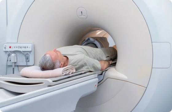
The patient lies on a table under a gamma camera, which detects gamma radiation and creates a detailed image of the heart. Photographs are taken when relaxed as well as stressed, caused by exercise or medication.
Nuclear Cardiology Stress Test
If exercise is part of the test, the patient will walk on a treadmill or ride a stationary bike to get the heart rate up. If the patient cannot exercise, medicines will be used to simulate the effects of exercise on the heart.
Second Image
After the stress test, second images are taken for comparison with
the resting images. This helps detect changes in blood flow to the heart muscle.
understanding the results and
what happens next
After your nuclear cardiology scan, the results are analyzed by a nuclear cardiologist who specializes in nuclear test cardiology. Findings will be discussed with the patient, highlighting key points, such as:
Presence of Coronary
Artery Disease
Identify areas of decreased blood flow that may indicate blockage in the coronary arteries.
Heart
Function
Assessment of how well the heart is beating and whether there is evidence of heart failure or decreased ejection fraction.
Tissue
Viability
Indicates that parts of the heart muscle are damaged and that they can benefit from revascularization.
Lifestyle
Modifications
Suggestions for diet, exercise, and other lifestyle changes to improve heart health.
Medication
Medications are prescribed or modified
to deal with conditions such as high blood pressure, high cholesterol, and heart failure.
Additional
Testing
In some cases, additional testing may be necessary to gather more information.
Surgical
Interventions
When severe blockages or other issues are detected, procedures such as angioplasty, stenting, or bypass surgery may be recommended.
why choose cardiology care for nuclear cardiology?
At Cardiology Care, we provide exceptional cardiovascular care using advanced technology and a patient-centered approach. Here’s why you should choose us for your nuclear cardiology in NYC:
Experienced Experts
We have a team of highly trained and experienced cardiologists who specialize in nuclear imaging and cardiac care.
Advanced Technology
We use the latest imaging technology to ensure accurate and precise diagnosis.
Comprehensive Care
We offer a simple and comprehensive care experience from the consultation to the follow-up care.


Individualized Treatment Plan
Each patient receives a customized treatment plan based on their specific health needs and risk factors.
Patient Education
We believe in empowering our patients with knowledge. We take the time to explain the procedure, side effects, and recommended treatment in detail.
frequently asked questions
What is a cardiac nuclear stress test?
Cardiac stress testing is a way to determine cardiac function and blood flow during physiological stress. It can combine exercise or medical stress with nuclear imaging to detect abnormal blood flow to the heart. A cardiac nuclear stress test involves injecting a radiotracer into the bloodstream and taking pictures of the heart at rest and after stress to give detailed information about how well the heart is working and whether the heart disease or low blood pressure.
What is a nuclear heart test?
A nuclear heart test, also known as a myocardial perfusion imaging (MPI) test, is a type of nuclear cardiology procedure used to examine the blood supply to the heart muscle. It involves inserting a radioactive probe and imaging with a gamma camera to determine the quality of blood flow to different parts of the heart. This test helps to diagnose pulmonary artery disease or stable ischemic heart disease, determine the severity of heart failure, and plan appropriate treatment strategies.
What is involved in a nuclear cardiac stress test?
The nuclear stress tests involve several steps:
Preparation
The patient is instructed to avoid eating, drinking, or taking certain medications for a few hours before the test.
Resting Images
The patient receives an injection of a radiotracer while at rest. After allowing time for the tracer to circulate, a gamma camera captures heart images.
Stress Induction
The patient undergoes physical exercise (usually on a treadmill or stationary bike) or receives a medication that mimics the effects of exercise on the heart. Also, the dobutamine stress test is used if you’re unable to exercise. This increases the heart’s workload and blood flow.
Nuclear Stress Test
Another injection of the radiotracer is given at peak exercise or after medication administration. The Regadenoson stress test is the most used. Images are retaken to assess enough blood flow during stress.
Comparison
The resting and stress images are compared to identify any areas with reduced blood flow or damage, indicating potential blockages or other heart issues. A nuclear stress test can diagnose coronary artery disease and show how severe the condition is.
The test provides valuable information on the heart’s blood supply, function, and overall health, aiding in diagnosing and managing cardiovascular conditions.


